
Optogenetics is a method that makes chosen types of nerve cells respond to light by giving them the gene for a light-sensitive protein such as channelrhodopsin, so that researchers can switch those cells on or off with millisecond precision [1, 2]. On 5 October 2026 the Nobel Assembly at Karolinska Institutet awarded the Nobel Prize in Physiology or Medicine jointly to Karl Deisseroth, Peter Hegemann and Georg Nagel “for their discoveries concerning light-gated ion channels and optogenetics” [3]. Our full report on the 2026 Nobel Prize in Physiology or Medicine covers the announcement and the three laureates.
For most of the twentieth century, researchers could link brain regions to behaviours but could not directly test causation [2]. Optogenetics changed that, because a scientist can now switch a defined population of neurons on or off and test whether those cells are sufficient or necessary for a behaviour [1].
Optogenetics: key facts
- Optogenetics places the gene for a light-gated ion channel into a chosen type of nerve cell, often with a virus, so that light delivered to those cells switches them on or off with millisecond timing (Nobel scientific background, PDF) [1].
- Channelrhodopsin-1, reported in Science on 28 June 2002 by Georg Nagel, Peter Hegemann, Ernst Bamberg and colleagues, is described by the Nobel Committee as the first direct evidence of a light-gated ion channel (PubMed 12089443) [4, 1].
- Channelrhodopsin-2 (ChR2), reported in PNAS on 25 November 2003 by the same team of Nagel, Hegemann, Bamberg and colleagues, is a directly light-gated channel for positive ions (cations) that opens under blue light (PubMed 14615590) [5, 1].
- Halorhodopsin (NpHR), added to the toolkit in 2007, is a light-driven pump that silences neurons under yellow light and allowed two-colour, bidirectional control alongside ChR2 (PubMed 17410168) [6, 1].
- The 2026 Nobel Prize in Physiology or Medicine, announced on 5 October 2026, went jointly to Karl Deisseroth (born 1971), Peter Hegemann (born 1954) and Georg Nagel (born 1953), who share 12 million Swedish kronor equally (Nobel press release, PDF) [3].
- In 2005, Edward Boyden, Feng Zhang, Ernst Bamberg, Georg Nagel and Karl Deisseroth made cultured rat hippocampal neurons carrying ChR2 fire on blue light with millisecond precision (PubMed 16116447) [7].
- In 2007, Deisseroth’s group drove whisker movements in living rodents through an implanted optical fibre, and a Deisseroth and de Lecea collaboration woke sleeping mice by stimulating hypocretin neurons with light (PubMed 17873414, 17943086) [8, 9].
- In 2021, Sahel and colleagues reported partial recovery of visual function in one blind patient with retinitis pigmentosa after optogenetic gene therapy encoding the opsin ChrimsonR and light-projecting goggles (PubMed 34031601) [10].
In Simple Words
A pond alga carries a protein that works like a tiny gate. When light hits the gate, it opens and lets electrically charged particles into the cell. Scientists copied the gene for that gate into one chosen kind of brain cell in laboratory animals. Now a flash of blue light makes only those cells fire. By watching what the animal does while the light is on, researchers learn what those cells are for. The three scientists who found the gate and turned it into a brain tool won the 2026 Nobel Prize [3, 2].
How Does Optogenetics Work?
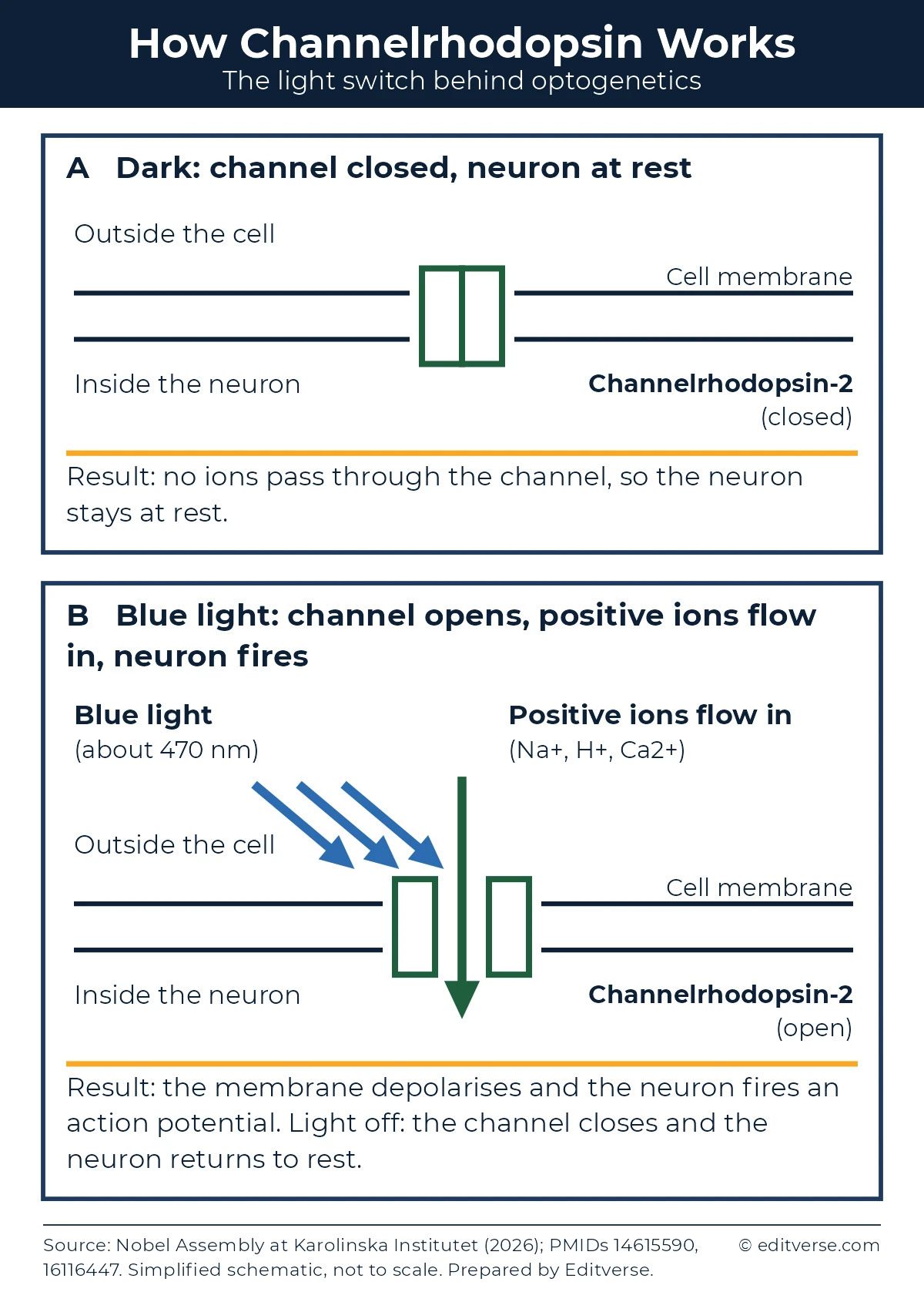
Optogenetics works by placing a light-gated ion channel in the membrane of chosen neurons, so that a pulse of light opens the channel, lets positive ions flow in, and makes the neuron fire [3, 5, 7].
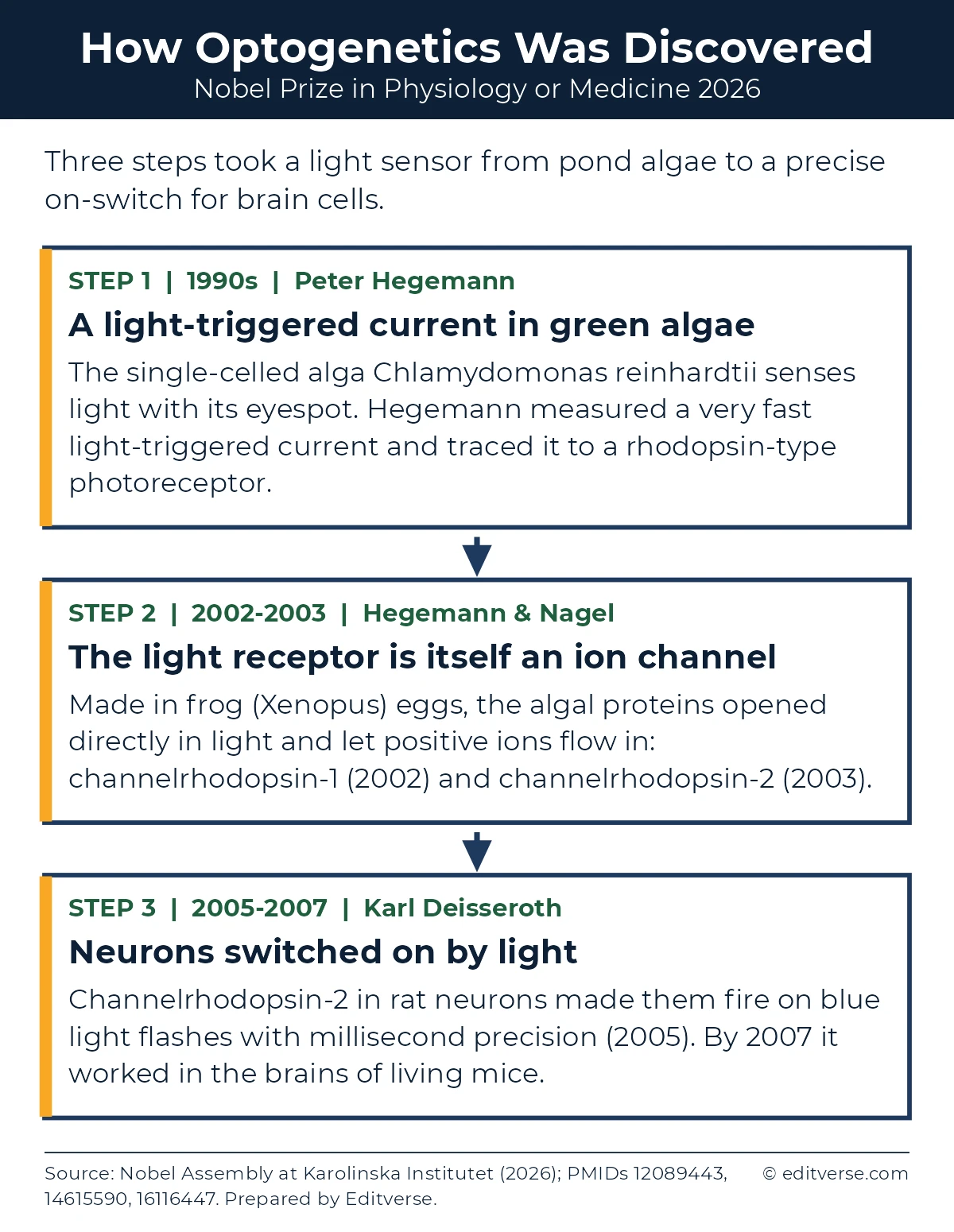
Chlamydomonas is a single-celled green alga that swims towards light, sensing it through an orange eyespot that contains the light-capturing molecule retinal [2]. Human vision relies on a multi-step cascade taking at least 10 ms, whereas the alga responds more than 20 times faster, which pointed to a single-protein system [2]. Channelrhodopsin turned out to be exactly that, one protein acting as both light sensor and ion channel [3, 2].
Channelrhodopsin-2, the switch that turns neurons on
When blue light strikes channelrhodopsin-2, a pore opens through the protein and positively charged ions flow into the cell [3, 5]. In frog egg cells the channel opened within 0.2 milliseconds of illumination [2]. Because ions flow passively through a channel rather than being pumped one at a time, the current is fast enough to match neuronal action potentials [1]. Inside a neuron, that inward current depolarises the membrane and triggers firing, and switching the light off closes the channel again.
ChR2 only switches neurons on. It cannot silence them. Mammalian neurons already contain enough retinal for the protein to work, so no chemical has to be added, an observation the Nobel Committee credits to Georg Nagel [1, 7].
Switching neurons off
Silencing requires different proteins. In 2007, Feng Zhang and colleagues in Deisseroth’s laboratory, working with Nagel and Gottschalk among others, added halorhodopsin (NpHR) from an archaeon, which suppresses firing under yellow light [6, 1]. Han and Boyden independently reached the same goal that year [1]. Because ChR2 responds to blue light and NpHR to yellow, both could be used in the same circuit for bidirectional control [1]. Pumps such as halorhodopsin move only about one ion per photon, however, and the discovery of natural anion channelrhodopsins in 2015 made silencing more efficient [1].
What a Typical Experiment Looks Like, Step by Step
A typical optogenetics experiment follows five steps, from choosing a cell type to watching how the animal or circuit responds when the light comes on [1, 7, 8]. Real protocols add controls at every stage.
- Pick the target cell type. Choose the population of neurons whose role you want to test, such as the hypocretin neurons studied in 2007 [9].
- Deliver the gene. Carry the channelrhodopsin-2 gene into the tissue, usually with a viral vector built so that only the chosen cell type expresses it. Boyden and colleagues used a lentivirus in cultured neurons in 2005 [7, 1].
- Let only the target cells respond. Genetic targeting means only the chosen neurons produce the light-sensitive channel, so neighbouring cells stay unaffected [1].
- Deliver light. Shine blue light onto the cells. In living rodents, Deisseroth’s group used a laser diode coupled to an optical fibre of about 200 micrometres inserted into the brain [8].
- Observe the effect. Record behaviour or circuit activity while switching the light on and off with millisecond timing, for example whisker deflections or transitions from sleep to wakefulness [8, 9].

Worked example: waking a sleeping mouse
The 2007 study by Adamantidis, de Lecea, Deisseroth and colleagues shows the logic of the method well. Hypocretin (orexin) neurons were already thought to support wakefulness, but nobody could say whether their activity caused awakening or merely accompanied it [1]. Researchers targeted ChR2 to these neurons, shone light on them in freely moving mice, and saw more transitions from sleep to wakefulness [9]. Recording alone could never provide that kind of sufficiency test [1].
Key Tools and Components
Every optogenetics experiment combines a light-sensitive protein, a way to get its gene into the right cells, a way to deliver light, and a way to read out the result [1, 2]. Each example below comes from the papers and Nobel documents cited here.
| Component | What it does | Examples from the literature | Source |
|---|---|---|---|
| Activating opsin | Opens under light, lets positive ions in, makes the neuron fire | Channelrhodopsin-2, activated by blue light | [5, 7] |
| Silencing opsin | Suppresses firing under light | Halorhodopsin NpHR (yellow light, 2007), anion channelrhodopsins (2015) | [6, 1] |
| Gene delivery | Carries the opsin gene into cells | Lentivirus in cultured rat neurons (2005), adeno-associated virus (AAV) in the human retina (2021) | [7, 10] |
| Genetic targeting | Restricts the opsin to one chosen cell type | ChR2 targeted to hypocretin neurons (2007), ChR2 targeted to excitatory layer-5 motor cortex neurons with the CaMKIIalpha promoter (2007) | [9, 8, 1] |
| Light delivery | Brings light of the right colour to the cells | Laser diode coupled to an optical fibre of about 200 micrometres (2007), light-projecting goggles (2021) | [8, 10] |
| Cofactor | Retinal, the light-capturing molecule the opsin needs | Natural levels in mammalian neurons are sufficient, so none is added | [1] |
| Readout | Measures what the light did | Spike recordings in culture, whisker deflection, sleep-to-wake transitions, fear-memory recall | [7, 8, 9, 11] |
The History of Optogenetics in Brief
Optogenetics grew from Peter Hegemann’s studies of how a green alga senses light, from Georg Nagel’s experiments with Hegemann and Ernst Bamberg showing that the algal light receptors are themselves ion channels, and from Karl Deisseroth’s use of channelrhodopsin-2 to control neurons [3, 1]. Credit belongs to a German and American collaboration, not to one laboratory.
Francis Crick argued in 1979 that neuroscience needed a way to control one cell type at a time, and in 1999 suggested that light would be the ideal signal, although he called the idea far-fetched [1, 2]. Older groundwork came from Oesterhelt and Stoeckenius, whose 1971 paper described bacteriorhodopsin and whose 1973 paper showed it is a light-driven proton pump [1]. In 1991, Harz and Hegemann recorded a light-evoked current in Chlamydomonas that began within a fraction of a millisecond [12, 2], and in the early 1990s he proposed, to considerable scepticism, that one protein both captured light and formed the channel [2].
A Chlamydomonas gene-sequence database released by Japanese researchers in 2000 made it possible to find the algal opsin genes [1, 2]. In 2002, John Spudich’s group used RNA interference to show that two rhodopsins drive the alga’s phototaxis [13], and Nagel, Hegemann, Bamberg and colleagues recorded light-gated currents from channelrhodopsin-1 in frog eggs [4]. Their 2003 PNAS paper, with Nagel as first author and Hegemann and Bamberg among the co-authors, characterised channelrhodopsin-2 and proposed it as a tool to depolarize cells simply by illumination [5, 1].
In 2005, Boyden, Zhang, Bamberg, Nagel and Deisseroth delivered ChR2 into cultured rat hippocampal neurons and drove spikes with millisecond precision [7]. Confirmations followed quickly, including work by Nagel, Gottschalk and colleagues showing light-triggered behaviour in the worm C. elegans [14]. The word “optogenetics” first appeared in print in a 2006 review by Deisseroth and five co-authors [15], light control of motor cortex in living rodents and the sleep-to-wake study followed in 2007 [8, 9], and Nature Methods named optogenetics its Method of the Year 2010 [16]. In 2013 the Brain Prize recognised six contributors, namely Ernst Bamberg, Edward Boyden, Karl Deisseroth, Peter Hegemann, Gero Miesenböck and Georg Nagel [17].
For a paper-by-paper account of the three founding publications, read our companion article on the papers behind the 2026 Nobel Prize in optogenetics.
What Has Optogenetics Been Used For?
Optogenetics has been used mainly to map which neural circuits control behaviours, memories and emotions in laboratory animals, and, in one reported patient, to partially restore vision [2, 1, 10].
- Circuit mapping. Researchers have identified circuits for pain, social behaviour, thirst, food intake, reward and attention, as well as cells controlling circadian rhythm and immune-triggered fever [2].
- Memory. In 2012, Liu, Ramirez, Deisseroth, Susumu Tonegawa and colleagues reactivated a hippocampal memory trace with light and triggered fear-memory recall in mice [11].
- Emotion and anxiety. Studies have separated reward from aversion in dopamine circuits and traced amygdala pathways for anxiety and fear [1].
- Innate behaviour. Aggression, mating and parenting have each been traced to distinct neural circuits [1, 2].
- Beyond the brain. Deisseroth showed that heart rhythm can influence anxiety, and other groups identified gut cells that help explain a preference for sugar over sweeteners [2].
- Disease biology. Optogenetics has improved understanding of depression, anxiety, schizophrenia, Alzheimer’s disease and Parkinson’s disease [2].
Optogenetics in humans: the 2021 vision case
The clinical milestone highlighted by the Nobel Committee involved a single patient. In 2021, Sahel, Roska and colleagues treated one blind person with retinitis pigmentosa, an inherited disease that destroys the eye’s rods and cones, by delivering the gene encoding the opsin ChrimsonR to the retina with an adeno-associated viral vector [10, 1]. With light-projecting goggles, the patient could discern and grasp objects on a table [2]. Its authors described it as the “first reported case of partial functional recovery in a neurodegenerative disease after optogenetic therapy” [10].
That result is a proof of concept in one patient, not a cure, and several clinical trials in retinitis pigmentosa are now under way [1, 2]. No optogenetic therapy for a brain disorder has been reported in the sources cited here.
Limitations and Open Questions
Optogenetics requires both genetic modification of the target cells and a way to deliver light to them, and those two requirements set most of its practical limits [1, 2].
- Gene delivery is a prerequisite. Each experiment depends on getting the opsin gene into the right cells, typically with a virus, which in humans means a gene-therapy step [1, 10].
- Light has to reach the cells. In the 2007 rodent experiments, light arrived through an optical fibre inserted into the brain [2, 8], an invasive arrangement.
- Silencing tools work differently from ChR2. Pumps such as halorhodopsin move about one ion per photon, which motivated the search for more efficient light-gated inhibitory channels [1].
- Human evidence is thin. One treated patient cannot establish how well a therapy works across a population, and trial results will decide that [10, 1].
As general guidance rather than a sourced finding, a clear methods section names the opsin, vector and light parameters and describes control conditions. Animal studies should also follow our guide to ARRIVE guidelines for animal research reporting and our advice on writing replicable methodology sections.
Optogenetics vs Other Neuromodulation Methods
Optogenetics differs from other neuromodulation methods by combining cell-type targeting with millisecond timing, whereas earlier methods could establish correlations but could not directly test whether specific cells contribute to a behaviour [1, 2].
| Method | How it acts | What the Nobel documents say | Source |
|---|---|---|---|
| Optogenetics | Light opens or closes genetically targeted ion channels or pumps | Gives millisecond control of defined cell types, so researchers can test cause and effect | [1] |
| Chemogenetics (DREADDs, Roth laboratory, 2007) | Engineered receptors respond to an otherwise inactive designer drug | Slower and complementary to optogenetics | [1] |
| Electrical stimulation (cochlear implants) | Electric current stimulates the auditory nerve | Optogenetic implants might activate the auditory nerve with greater precision | [2] |
Optical methods for imaging as well as stimulating the brain are covered in our explainer on neurophotonics and optical brain research.
Professional Support for Neuroscience Manuscripts
The three founding papers appeared in Science, PNAS and Nature Neuroscience. For your own neuroscience manuscript, our guide on how to publish in top neuroscience journals sets out what editors expect. Four services suit methods-heavy neuroscience papers best.
Substantive Editing
$250
Subject-expert editing for clarity, flow and formatting, with unlimited revisions.
Journal Selection
$115
Five to ten ranked journal suggestions with a rationale covering scope, impact factor and acceptance rate.
Results and Discussion Writing
$400
A detailed Results and Discussion section with citations. 4 days.
Graphical Abstract
$150
A journal-sized graphical abstract for your study. 2-3 days.
Aiming higher? Read our article on publishing in Nature and other high-impact multidisciplinary journals.
Free Research Tools from Editverse
- GraphPro: build publication-ready charts, 25 types, with built-in t-test, ANOVA, correlation, and chi-square p-values. Free, no signup.
- StatPro: free statistical calculators and a test-selector tool covering group comparisons, categorical outcomes, time-to-event analysis, and diagnostic accuracy.
- Flowchart Pro: generate PRISMA 2020, PRISMA-ScR, CONSORT, STROBE, STARD, MOOSE, and ARRIVE flow diagrams directly in your browser.
Related Editverse Resources
- Nobel Prize in Medicine 2026: optogenetics and the three laureates
- The papers behind the 2026 Nobel Prize in optogenetics
- Neurophotonics: illuminating brain function
- How to publish in top neuroscience journals
- Exploring neuroscience and brain research advances
- ARRIVE guidelines for animal research reporting
- Writing clear, replicable methodology sections
- Publishing in Nature and other high-impact journals
Frequently Asked Questions About Optogenetics
What is optogenetics?
Optogenetics is a method that makes chosen types of nerve cells respond to light by giving them the gene for a light-sensitive protein such as channelrhodopsin, so that researchers can switch those cells on or off with millisecond precision. Light is delivered to the cells, for example through an implanted optical fibre, and the cells respond within milliseconds [1, 2].
How does optogenetics work?
Researchers deliver the gene for a light-gated ion channel such as channelrhodopsin-2 into a chosen type of neuron, often with a virus. When blue light reaches those cells, the channel opens, positive ions flow in, and the neuron fires. Silencing uses other proteins such as halorhodopsin [1, 5, 6].
Who invented optogenetics?
Optogenetics came from several groups. Peter Hegemann studied the light response of the alga Chlamydomonas, Georg Nagel, Hegemann and Ernst Bamberg showed in 2002 and 2003 that channelrhodopsins are light-gated ion channels, and Karl Deisseroth, Edward Boyden, Feng Zhang, Bamberg and Nagel used channelrhodopsin-2 to control neurons in 2005. Deisseroth, Hegemann and Nagel shared the 2026 Nobel Prize [3, 4, 5, 7].
Is optogenetics used in humans?
Only experimentally. In 2021 one blind patient with retinitis pigmentosa partially recovered visual function after optogenetic therapy combined with light-projecting goggles, and several clinical trials for this disease are under way. There is no approved optogenetic treatment in the sources cited here [10, 1].
What is channelrhodopsin-2?
Channelrhodopsin-2 (ChR2) is a protein from the green alga Chlamydomonas that acts as both a light sensor and an ion channel. Described by Nagel, Hegemann, Bamberg and colleagues in PNAS in November 2003, it opens under blue light and lets positive ions into the cell [5, 3].
What light is used in optogenetics?
Colour depends on the protein. Channelrhodopsin-2 is activated by blue light and halorhodopsin silences neurons under yellow light, while the 2021 vision therapy used a different opsin, ChrimsonR. Light reaches the cells through optical fibres in animal studies or light-projecting goggles in the 2021 vision study [5, 6, 2, 10].
Why did optogenetics win the Nobel Prize?
In its citation, the Nobel Assembly named the laureates’ discoveries concerning light-gated ion channels and optogenetics. Optogenetics lets researchers show how specific nerve cells shape memories, feelings and behaviours in the living brain, moving neuroscience from observing correlations to testing causes [3, 1].
Last verified: 7 October 2026. Updated 7 October 2026 to reflect the 2026 Nobel Prize in Physiology or Medicine. This version replaces an earlier article that contained unsourced quotations and historical errors.
How to cite this article
Editverse. (2026, October 7). Optogenetics: How light controls the brain (updated for the 2026 Nobel Prize). Editverse. https://editverse.com/optogenetics-controlling-the-brain-with-light/
References
- Nobel Committee for Physiology or Medicine. Scientific background 2026: Optogenetics, discovery of a neuronal switch [Internet]. Stockholm: Nobel Assembly at Karolinska Institutet; 2026 Oct 5 [cited 2026 Oct 7]. Available from: https://www.nobelprize.org/uploads/2026/10/advanced-medicineprize2026.pdf
- Nobel Assembly at Karolinska Institutet. Popular science background: A light-sensitive algal protein energised neuroscience [Internet]. Stockholm: Karolinska Institutet; 2026 Oct 5 [cited 2026 Oct 7]. Available from: https://www.nobelprize.org/uploads/2026/10/popular-medicineprize2026.pdf
- Nobel Assembly at Karolinska Institutet. Press release: The 2026 Nobel Prize in Physiology or Medicine [Internet]. Stockholm: Karolinska Institutet; 2026 Oct 5 [cited 2026 Oct 7]. Available from: https://www.nobelprize.org/uploads/2026/10/press-medicineprize2026.pdf
- Nagel G, Ollig D, Fuhrmann M, Kateriya S, Musti AM, Bamberg E, et al. Channelrhodopsin-1: a light-gated proton channel in green algae. Science. 2002 Jun 28;296(5577):2395-8. doi: 10.1126/science.1072068. PMID: 12089443
- Nagel G, Szellas T, Huhn W, Kateriya S, Adeishvili N, Berthold P, et al. Channelrhodopsin-2, a directly light-gated cation-selective membrane channel. Proc Natl Acad Sci U S A. 2003 Nov 25;100(24):13940-5. doi: 10.1073/pnas.1936192100. PMID: 14615590
- Zhang F, Wang LP, Brauner M, Liewald JF, Kay K, Watzke N, et al. Multimodal fast optical interrogation of neural circuitry. Nature. 2007 Apr 5;446(7136):633-9. doi: 10.1038/nature05744. PMID: 17410168
- Boyden ES, Zhang F, Bamberg E, Nagel G, Deisseroth K. Millisecond-timescale, genetically targeted optical control of neural activity. Nat Neurosci. 2005 Sep;8(9):1263-8. doi: 10.1038/nn1525. PMID: 16116447
- Aravanis AM, Wang LP, Zhang F, Meltzer LA, Mogri MZ, Schneider MB, et al. An optical neural interface: in vivo control of rodent motor cortex with integrated fiberoptic and optogenetic technology. J Neural Eng. 2007 Sep;4(3):S143-56. doi: 10.1088/1741-2560/4/3/S02. PMID: 17873414
- Adamantidis AR, Zhang F, Aravanis AM, Deisseroth K, de Lecea L. Neural substrates of awakening probed with optogenetic control of hypocretin neurons. Nature. 2007 Nov 15;450(7168):420-4. doi: 10.1038/nature06310. PMID: 17943086
- Sahel JA, Boulanger-Scemama E, Pagot C, Arleo A, Galluppi F, Martel JN, et al. Partial recovery of visual function in a blind patient after optogenetic therapy. Nat Med. 2021 Jul;27(7):1223-9. doi: 10.1038/s41591-021-01351-4. PMID: 34031601
- Liu X, Ramirez S, Pang PT, Puryear CB, Govindarajan A, Deisseroth K, et al. Optogenetic stimulation of a hippocampal engram activates fear memory recall. Nature. 2012 Mar 22;484(7394):381-5. doi: 10.1038/nature11028. PMID: 22441246
- Harz H, Hegemann P. Rhodopsin-regulated calcium currents in Chlamydomonas. Nature. 1991 Jun;351(6326):489-91. doi: 10.1038/351489a0
- Sineshchekov OA, Jung KH, Spudich JL. Two rhodopsins mediate phototaxis to low- and high-intensity light in Chlamydomonas reinhardtii. Proc Natl Acad Sci U S A. 2002 Jun 25;99(13):8689-94. doi: 10.1073/pnas.122243399. PMID: 12060707
- Nagel G, Brauner M, Liewald JF, Adeishvili N, Bamberg E, Gottschalk A. Light activation of channelrhodopsin-2 in excitable cells of Caenorhabditis elegans triggers rapid behavioral responses. Curr Biol. 2005 Dec 20;15(24):2279-84. doi: 10.1016/j.cub.2005.11.032. PMID: 16360690
- Deisseroth K, Feng G, Majewska AK, Miesenböck G, Ting A, Schnitzer MJ. Next-generation optical technologies for illuminating genetically targeted brain circuits. J Neurosci. 2006 Oct 11;26(41):10380-6. doi: 10.1523/JNEUROSCI.3863-06.2006. PMID: 17035522
- Method of the Year 2010 [editorial]. Nat Methods. 2011 Jan;8(1):1. Published online 2010 Dec 20. doi: 10.1038/nmeth.f.321
- Federation of European Neuroscience Societies. Six eminent scientists share the world’s largest brain research prize [Internet]. FENS; 2013 [cited 2026 Oct 7]. Available from: https://fens.org/news-activities/news/six-eminent-scientists-share-the-worlds-largest-brain-research-prize