“The true method of discovery is like the flight of an aeroplane. It starts from the ground of particular observation; it makes a flight in the thin air of imaginative generalization; and it again lands for renewed observation rendered acute by rational interpretation.” – Alfred North Whitehead
Radiomics: Mining Medical Images for Insights
Radiomics is an emerging field that combines advanced image analysis with data mining techniques to extract quantitative features from medical images. This guide explores how radiomics is revolutionizing medical diagnosis, treatment planning, and prognosis prediction by unlocking hidden information within imaging data.
“Radiomics has the potential to uncover disease characteristics that fail to be appreciated by the naked eye, offering a powerful tool for personalized medicine.”
— Dr. Robert J. Gillies, Pioneer in Radiomics Research
1. Understanding Radiomics
Radiomics is a data-driven approach to medical imaging analysis:
Key Concepts:
- Definition: The high-throughput extraction of quantitative features from medical images
- Imaging Modalities: CT, MRI, PET, ultrasound, and more
- Feature Types: Shape, intensity, texture, and higher-order statistics
- Data Mining: Machine learning and statistical analysis of extracted features
- Integration: Combining radiomics data with other patient information (genomics, clinical data)
2. The Radiomics Workflow
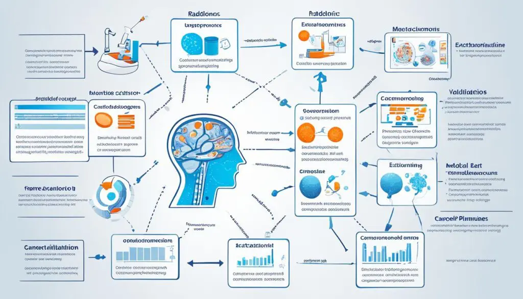
The process of radiomics analysis involves several key steps:
Workflow Stages:
- Image Acquisition: Obtaining high-quality medical images
- Image Segmentation: Delineating regions of interest (ROIs)
- Feature Extraction: Calculating quantitative features from ROIs
- Feature Selection: Identifying the most relevant features
- Model Building: Developing predictive or diagnostic models
- Validation: Testing model performance on independent datasets
- Clinical Application: Integrating radiomics models into clinical decision-making
3. Applications in Oncology
Radiomics has shown significant promise in cancer research and treatment:
Oncological Applications:
- Tumor Characterization: Non-invasive assessment of tumor heterogeneity and aggressiveness
- Treatment Response Prediction: Identifying patients likely to respond to specific therapies
- Prognosis Estimation: Predicting patient outcomes and survival rates
- Early Detection: Improving screening and early diagnosis of cancers
- Precision Medicine: Tailoring treatment plans based on individual tumor characteristics
- Recurrence Monitoring: Detecting early signs of cancer recurrence
4. Beyond Oncology
Radiomics is expanding into other medical domains:
Emerging Applications:
- Neurology: Analyzing brain structure and function in neurological disorders
- Cardiology: Assessing cardiac function and predicting cardiovascular events
- Pulmonology: Characterizing lung diseases and predicting outcomes
- Musculoskeletal Imaging: Evaluating bone and joint conditions
- Hepatology: Assessing liver diseases and fibrosis stages
- Ophthalmology: Analyzing retinal images for disease detection
5. Advanced Techniques in Radiomics
Recent advancements are pushing the boundaries of radiomics:
Cutting-edge Approaches:
- Deep Learning Radiomics: Using convolutional neural networks for feature extraction
- Delta-radiomics: Analyzing changes in radiomic features over time
- Radiogenomics: Correlating imaging features with genomic profiles
- Multi-modal Radiomics: Integrating features from different imaging modalities
- Habitat Radiomics: Analyzing tumor sub-regions or “habitats”
- Radiomics Nomograms: Combining radiomics with clinical factors for decision support
6. Challenges in Radiomics
Despite its potential, radiomics faces several challenges:
Key Challenges:
- Standardization: Ensuring consistency in image acquisition and feature extraction
- Reproducibility: Validating results across different centers and datasets
- Feature Selection: Identifying the most relevant features among thousands
- Biological Interpretation: Understanding the biological meaning of radiomic features
- Data Quality: Dealing with noise, artifacts, and variability in medical images
- Clinical Integration: Incorporating radiomics into clinical workflows
7. Ethical and Legal Considerations
The use of radiomics raises important ethical and legal questions:
Key Considerations:
- Data Privacy: Protecting patient information in large-scale radiomics studies
- Informed Consent: Ensuring patients understand the use of their imaging data
- Algorithm Transparency: Explaining radiomics models to patients and clinicians
- Regulatory Approval: Navigating the approval process for radiomics-based tools
- Liability: Determining responsibility for errors in radiomics-guided decisions
- Health Disparities: Addressing potential biases in radiomics models
8. Future Directions
The field of radiomics is rapidly evolving:
Emerging Trends:
- Federated Learning: Collaborative model training across institutions without data sharing
- Explainable AI: Developing interpretable radiomics models
- Real-time Radiomics: Integrating radiomics analysis into live imaging workflows
- Radiomics-guided Interventions: Using radiomics for treatment planning and guidance
- Liquid Biopsy Integration: Combining radiomics with circulating biomarkers
- Synthetic Data Generation: Creating artificial datasets for model training and validation
9. Tools and Resources
Various tools and resources are available for radiomics research:
Radiomics Resources:
- Software Packages: PyRadiomics, IBEX, LifeX, RaCaT
- Image Databases: The Cancer Imaging Archive (TCIA), UK Biobank Imaging
- Standardization Initiatives: Image Biomarker Standardisation Initiative (IBSI)
- Reporting Guidelines: Radiomics Quality Score (RQS)
- Conferences: MICCAI, SPIE Medical Imaging, RSNA
- Journals: Radiology: Artificial Intelligence, Medical Physics, Scientific Reports
10. Interdisciplinary Collaboration
Advancing radiomics requires collaboration across disciplines:
Key Collaborations:
- Radiology: Expertise in medical imaging and interpretation
- Computer Science: Development of algorithms and machine learning models
- Biostatistics: Design of studies and statistical analysis of radiomics data
- Oncology: Clinical expertise in cancer diagnosis and treatment
- Bioinformatics: Integration of radiomics with genomic and molecular data
- Medical Physics: Understanding of image acquisition and processing
Conclusion
Radiomics represents a paradigm shift in medical imaging, transforming the way we extract and analyze information from medical images. By harnessing the power of advanced computational techniques, radiomics has the potential to:
- Enhance diagnostic accuracy and precision
- Personalize treatment strategies
- Improve patient outcomes through early detection and intervention
- Accelerate drug discovery and development
- Provide non-invasive alternatives to traditional diagnostic methods
As the field continues to evolve, addressing challenges such as standardization, reproducibility, and clinical integration will be crucial. The integration of radiomics with other emerging technologies, such as artificial intelligence and multi-omics approaches, promises to further revolutionize healthcare delivery.
The future of radiomics lies in its ability to unlock the wealth of information hidden within medical images, offering a powerful tool for precision medicine and paving the way for more effective, personalized healthcare strategies. As research progresses and clinical applications expand, radiomics is poised to play an increasingly vital role in shaping the future of medicine.
“Radiomics is not just about extracting more data from images; it’s about uncovering the subtle patterns and relationships that can transform our understanding of diseases and guide personalized treatment decisions.”
— Dr. Hugo Aerts, Pioneer in Radiomics Research
In the fast-changing world of healthcare, radiomics is a key innovation. It uses advanced computer analysis to find hidden insights in medical scans. By mining medical images, radiomics helps doctors and researchers. They can see tumor types, predict treatment success, and lead to personalized medicine.
This new way of looking at medical images started with visionaries who saw their huge potential. Now, radiomics is growing fast. It’s bringing new ways to understand Medical Image Phenotyping, Radiogenomics, and combining Radiomic Features with Machine Learning in Radiology.

Key Takeaways
- Radiomics uses advanced computer analysis to find valuable insights in medical images.
- It helps discover tumor types, predict treatment outcomes, and supports personalized medicine.
- Radiomics comes from Quantitative Image Analysis, Biomedical Imaging Informatics, and Computational Radiology.
- Its applications include Medical Image Phenotyping, Radiogenomics, and combining Radiomic Features with Machine Learning.
- Radiomics is changing healthcare by unlocking the full potential of medical imaging data.
Introduction to Radiomics
Radiomics is changing how we look at medical scans. It uses advanced algorithms to find hidden info in images. This method goes way beyond what our eyes can see.
What is Radiomics?
Radiomics pulls out lots of details from scans like CT, MRI, and PET. These details help us understand diseases better, especially in oncology.
The Evolution of Medical Imaging and Quantitative Analysis
Medical imaging has come a long way from X-rays to today’s high-tech scans. At the same time, analyzing images has gotten more advanced. Radiomics uses these new techs to change how we diagnose and treat diseases.
“Radiomics has the ability to identify tumor genotypes and pathological imaging biomarkers with clinical potential.”
Radiomics: A Powerful Tool for Oncology
Radiomics is a key tool in oncology. It helps by looking at many features in medical images. This lets us understand tumors better, including their biology and genetics.
Decoding Tumor Phenotypes
Radiomics can show us the detailed features of tumors. It uses advanced image analysis to find things like texture and shape. These details help match with clinical and genetic data, giving us a deeper look at the tumor.
Predicting Treatment Outcomes
Radiomics can also predict how well treatments will work. This means doctors can choose the best treatment for each patient. It helps predict things like treatment success, survival rates, and the chance of the disease coming back.
Adding radiomics to cancer care could change how we manage cancer. It gives doctors valuable insights for making treatment plans. As radiomics grows, it will likely play a big part in improving cancer care and enhancing clinical decision-making.
“Radiomics, the high-throughput mining of quantitative image features from medical imaging, is gaining importance in cancer research.”
Radiomics Workflow and Challenges
The radiomics workflow includes key steps like image capture, segmenting, feature pulling, and analyzing data. Each step has its own hurdles that need solving for radiomics to work well in clinics.
One big challenge is making sure imaging methods are the same everywhere. Things like slice thickness and how the images are made can change the results. To fix this, researchers aim to create imaging methods that work the same everywhere.
Getting the tumor area right through segmentation is also tough. It’s key for getting accurate features and analysis. But, doing it by hand takes a lot of time and can vary a lot from person to person. Using automated methods, like deep learning, looks promising but needs lots of good data to work right.
- There are so many features in radiomics that it can overfit, meaning it works great on the data it knows but not on new data. To fix this, we need to pick the best features and reduce the number of them, and make sure they’re tested well.
- Combining radiomic data with other health info is key for making radiomics useful in treating patients as individuals. But, this is hard because of the need to make all the data work together, handle missing data, and create models that predict well.
Even with these hurdles, the radiomics workflow is getting better. Researchers are tackling the issues head-on. By focusing on making things standard, getting the tumor area right, choosing the right features, and combining data well, radiomics is set to bring big advances in diagnosing diseases, planning treatments, and improving patient care.

“Radiomics features are advanced through sophisticated mathematical analysis, aiming to enhance radiographic data in medical imaging.”
| Metric | Value |
|---|---|
| Article Accesses | 6437 |
| Citation Count | 31 |
| Altmetric Score | 13 |
By tackling the radiomics challenges and making the radiomics workflow smoother, researchers and doctors can make the most of this powerful tool. This will lead to better patient care and more tailored healthcare solutions.
Radiomic Features and Machine Learning
Radiomics is a big deal in precision medicine. It’s all about pulling and analyzing numbers from medical images. At the heart are radiomic features. These are many metrics that show the details of tumors and other body parts. They include things like intensity, texture, and shape.
Machine learning has changed the game for radiomics. These algorithms help pick the best features and make predictions. This leads to better diagnosis, treatment plans, and patient care.
Extracting Radiomic Features
Getting radiomic features is key. Researchers use many methods to look at medical images. They look at:
- First-order statistics: This is about the distribution of pixel intensities, like the average and how spread out they are.
- Textural features: This looks at how pixels relate to each other, showing patterns and differences in the image.
- Shape-based features: These capture the size, shape, and other details of the area being studied.
Machine Learning in Radiomics
Machine learning has sped up radiomics. It finds the most useful features and makes predictions. Researchers use different methods, like:
- Supervised learning: This uses labeled data to train models for tasks like predicting treatment success or disease types.
- Unsupervised learning: This finds patterns in data without labels, helping discover new insights and markers.
- Ensemble methods: This combines models to get better predictions and make radiomics more reliable.
Putting radiomics and machine learning together has changed healthcare. It leads to more accurate and tailored medical decisions. As this field grows, these tools will keep playing a big part in precision medicine.
Radiomics Applications in Clinical Practice
Radiomics is a powerful way to find valuable insights in medical images. It’s especially useful in oncology. It looks at how imaging features relate to a tumor’s genes or molecules.
Radiogenomics
Radiomics and genomics together help doctors understand tumors better. This leads to treatments made just for the patient. Researchers have found links between imaging and a tumor’s genes, helping in cancer care decisions.
Personalized Medicine
Radiomics helps move towards personalized medicine. It looks at each patient’s tumor in detail. This leads to treatments that fit the patient’s needs, improving health outcomes and patient happiness.
The study of radiomics applications is growing fast, about 178% a year. Radiogenomics and personalized medicine are changing how we fight cancer. Radiomics is key in making these changes, leading to better cancer care.
“Radiomics offers advantages over traditional methods by utilizing existing medical images, eliminating the need for invasive procedures, reducing patient discomfort, risk of complications, and healthcare costs associated with repeated imaging examinations.”
Radiomics and Precision Medicine Initiative
The Precision Medicine Initiative aims to change healthcare by making treatments fit each person’s needs. Radiomics is key to this goal. It lets doctors see and track a patient’s tumor without surgery. By using radiomics with the Precision Medicine Initiative, doctors can make healthcare more personal and effective.
Many studies show how radiomics and the Precision Medicine Initiative work together. A 2011 report talked about creating a new way to understand diseases for research. This led to using radiomics in healthcare. In 2015, the European Society of Radiology talked about how medical images help in making treatments more personal.
Articles in Radiology, Nature Reviews Clinical Oncology, and PLoS ONE have looked into radiomics and personalized medicine. They’ve used radiogenomics to understand diseases better. They used big databases like The Cancer Genome Atlas and The Cancer Imaging Archive.
Even though making radiomics work well is hard, it’s showing big promises. It could change how we treat complex diseases. As it grows, radiomics will likely play a big part in the Precision Medicine Initiative. This could lead to better healthcare for everyone.
“Radiomics aligns perfectly with the vision of the Precision Medicine Initiative, as it provides a non-invasive means of assessing and monitoring a patient’s tumor characteristics.”
Radiomics: Mining Medical Images for Insights
At the core of radiomics is the idea of mining medical images for valuable insights. Advanced computational methods are used on standard medical scans. This reveals information that would be missed otherwise. It helps in identifying tumor characteristics, predicting treatment outcomes, and uncovering genetic signatures.
Radiomics is great at spotting subtle image features that are crucial for understanding a patient’s health. With advanced algorithms and machine learning, it finds patterns in medical images that humans can’t see. This could change how we diagnose, monitor, and treat many diseases, from cancer to neurodegenerative disorders.
“Radiomics has gained significant traction and attention in recent years for its ability to identify tumor genotypes and pathological imaging biomarkers.”
The field of radiomics is growing fast, leading to more advanced uses in healthcare. It will help in making treatment plans tailored to each patient and in detecting diseases early. The insights from radiomics will be key in pushing healthcare forward.
The Future of Radiomics
The future of radiomics is thrilling. As new imaging and computational methods come out, we’ll get more detailed insights from medical images. By using radiomics, we can understand human health and disease better. This will lead to better health outcomes for people all over the world.
Radiomics Software and Tools
The radiomics workflow uses special software and tools. These help in getting, analyzing, and understanding radiomic features. There are both open-source and commercial options. Each has its own strengths and abilities.
Open-Source Radiomics Solutions
Open-source platforms like PyRadiomics and LifeX give users flexibility and customization. They let researchers and doctors explore radiomics deeply. Users can see and change the algorithms and methods used.
Commercial Radiomics Solutions
Commercial software, however, has more features and is easier to use. It also has advanced analytics and works well with other clinical data. These platforms might cost more but are great for those new to radiomics.
These software and tools have helped make radiomics more popular in medical imaging and oncology.
“The impact of quantitative imaging in medicine and surgery is a crucial element in charting the course for the future.”
– Wang Y-XJ, Ng CK, Quant Imaging Med Surg 2011
Future Directions and Challenges
As radiomics grows, we see many exciting new paths ahead. Improvements in imaging tech, machine learning, and computing will make radiomics more precise and useful. Combining radiomics with other fields like genomics and proteomics could lead to a deeper understanding of tumors and better treatments.
But, radiomics also faces big challenges. We need standard rules, solid checks, and easy use in hospitals. Overcoming these hurdles is key to making radiomics a big part of precision medicine.
Advancing Radiomics with Deep Learning
Deep Learning (DL) is a big win for radiomics, helping with precision medicine by spotting patient types and predicting results. CNNs are top picks for their great pattern spotting in medical images. Yet, sharing sensitive medical info online is risky, even with top-notch encryption. We must find ways to keep data safe while using DL in radiomics, even in places we can’t fully trust.
Ensuring Privacy in Radiomics
Studies have looked into how to keep medical info safe with Privacy-Preserving Machine Learning (PPML). They found we need better ways to organize and study these privacy tools for Deep Radiomics. These tools are key for keeping data safe, especially in Deep Radiomics where keeping things accurate, efficient, and private is a must.
| Radiomics Future Directions | Radiomics Challenges |
|---|---|
|
|

“Addressing the challenges in radiomics will be crucial for realizing its full potential in transforming healthcare.”
Conclusion
Radiomics is changing healthcare by using medical imaging data in new ways. It looks at lots of features in medical scans to understand tumors and how treatments work. This helps doctors make better choices for each patient.
Studies show how important radiomics is becoming. For example, over half of the research uses it to help with brain disorders. In cancer research, many studies look at how different tumors are and how they respond to treatments.
The future looks bright for radiomics. With better AI, radiomics will get even more powerful. This will help doctors give patients treatments that fit their unique needs. It’s a big step towards making healthcare more personal and effective.
FAQ
What is radiomics?
Radiomics is a way to analyze medical images deeply. It uses advanced algorithms to find and study many features in images. This helps understand tumors better, predict treatment success, and make treatments more personal.
How does radiomics evolve from the advancements in medical imaging and quantitative analysis?
Medical imaging has grown from simple X-rays to today’s high-tech scans like CT and MRI. At the same time, analyzing these images has become more advanced. This lets experts find new insights in medical scans.
What are the key applications of radiomics in oncology?
In oncology, radiomics is a key tool. It helps by looking at many features in medical images. This gives insights into tumor biology and can predict how well treatments will work.
What are the key steps in the radiomics workflow?
The radiomics process has several important steps. These include getting the images, segmenting them, finding features, and analyzing the data. Each step is crucial for making radiomics useful in real healthcare.
How do radiomic features and machine learning contribute to the radiomics workflow?
Radiomics looks at many features in medical images, like intensity and texture. Machine learning helps find the most useful features. This helps make predictions and develop new models.
What are the clinical applications of radiomics beyond oncology?
Radiomics is useful in many areas of healthcare, especially in oncology. It helps with radiogenomics, which links imaging with tumor genetics. It also supports personalized medicine by tailoring treatments to each patient’s tumor.
How does radiomics align with the Precision Medicine Initiative?
Radiomics fits well with the Precision Medicine Initiative. It offers a way to look at tumors without surgery. This helps make healthcare more personalized and effective.
What software and tools are available for radiomics?
There are many software and tools for radiomics. Some are open-source, others are commercial. These tools help with analyzing and understanding radiomic features, making radiomics more useful.
What are the future directions and challenges for radiomics?
Radiomics is growing fast, with many new areas to explore. It will need better imaging tech, smarter algorithms, and more computing power. But, it also faces challenges like standardizing methods and fitting into healthcare. Overcoming these will help make radiomics a big change in healthcare.
Source Links
- https://www.ncbi.nlm.nih.gov/pmc/articles/PMC7362913/ – Radiomics: from qualitative to quantitative imaging
- https://www.ncbi.nlm.nih.gov/pmc/articles/PMC4734157/ – Radiomics: Images Are More than Pictures, They Are Data
- https://www.mdpi.com/2073-8994/15/10/1834 – Radiomics and Its Feature Selection: A Review
- https://insightsimaging.springeropen.com/articles/10.1186/s13244-023-01437-2 – An overview of meta-analyses on radiomics: more evidence is needed to support clinical translation – Insights into Imaging
- https://www.mdpi.com/2072-6694/14/19/4871 – Oncologic Imaging and Radiomics: A Walkthrough Review of Methodological Challenges
- https://cris.maastrichtuniversity.nl/en/publications/radiomics-the-bridge-between-medical-imaging-and-personalized-med – Radiomics: the bridge between medical imaging and personalized medicine
- https://mmrjournal.biomedcentral.com/articles/10.1186/s40779-023-00458-8 – Artificial intelligence-driven radiomics study in cancer: the role of feature engineering and modeling – Military Medical Research
- https://www.termedia.pl/The-role-of-radiomics-in-dentistry-and-oral-r-nradiology,137,54186,1,1.html – The role of radiomics in dentistry and oral radiology
- https://www.ctisus.com/learning/pearls/3d-and-workflow/radiomics – 3D and Workflow: Radiomics Imaging Pearls – Educational Tools | CT Scanning | CT Imaging | CT Scan Protocols
- https://www.frontiersin.org/journals/oncology/articles/10.3389/fonc.2015.00272/full – Frontiers | Radiomic Machine-Learning Classifiers for Prognostic Biomarkers of Head and Neck Cancer
- https://www.appliedclinicaltrialsonline.com/view/radiomics-in-action – Radiomics in Action
- https://www.ncbi.nlm.nih.gov/pmc/articles/PMC8891653/ – Deep Learning With Radiomics for Disease Diagnosis and Treatment: Challenges and Potential
- https://www.mdpi.com/2075-4426/12/9/1373 – Is Radiomics Growing towards Clinical Practice?
- https://systems.enpress-publisher.com/index.php/MIPT/article/viewFile/6279/3022 – PDF
- https://www.nature.com/articles/s41571-022-00707-0 – Criteria for the translation of radiomics into clinically useful tests – Nature Reviews Clinical Oncology
- https://www.ncbi.nlm.nih.gov/pmc/articles/PMC9692256/ – Precision Medicine in Radiomics and Radiogenomics
- https://www.mdpi.com/2075-4426/12/11/1806?type=check_update&version=1 – Precision Medicine in Radiomics and Radiogenomics
- https://radiomics.bio/publications/publications/ – Publications I Radiomics
- https://www.linkedin.com/pulse/demystifying-black-box-ai-radiomics-methods-david-cain – Trustworthy AI: Building Confidence in Radiomic Analysis
- https://arxiv.org/html/2407.00538v1 – Privacy-Preserving and Trustworthy
Deep Learning for Medical Imaging ††thanks: This work has been submitted to the IEEE for possible publication. Copyright may be transferred without notice, after which this version may no longer be accessible. - https://www.scienceopen.com/hosted-document?doi=10.15212/RADSCI-2023-0018 – Large scale models in radiology: revolutionizing the future of medical imaging
- https://www.ncbi.nlm.nih.gov/pmc/articles/PMC8580417/ – AI in Medical Imaging Informatics: Current Challenges and Future Directions
- https://www.ncbi.nlm.nih.gov/pmc/articles/PMC7151556/ – From Medical Imaging to Radiomics: Role of Data Science for Advancing Precision Health
- https://www.linkedin.com/pulse/ai-radiomics-upgraded-medical-imaging-david-cain-l0x1c – AI and Radiomics: Precision Medical Imaging
