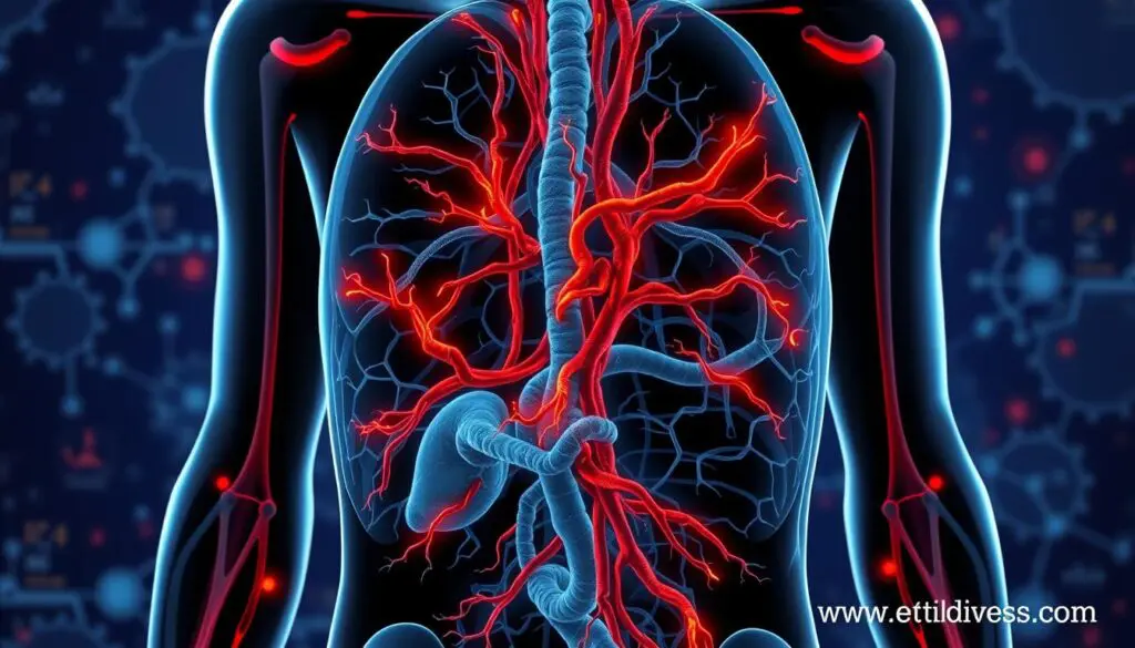
Giant cell arteritis (GCA) affects over 200,000 Americans yearly. It’s most common in older people of North European descent. Diagnosing it early is hard, leading to delayed treatment and serious issues like blindness or stroke. But, PET/CT imaging has changed how doctors find and treat vasculitis. It shows inflamed vessels clearly, helping doctors fight autoimmune diseases.
Key Takeaways
- PET/CT imaging can accurately identify inflammation in the arterial walls, helping distinguish vasculitis from atherosclerosis.
- PET/CT has demonstrated high sensitivity (77-92%) and specificity (89-100%) in detecting large vessel vasculitis, particularly in atypical or complex cases.
- PET/CT allows for early diagnosis of vasculitis involving the extratemporal proximal vessels, where traditional temporal artery biopsies may be non-diagnostic.
- Advancements in PET imaging, including digital long axial field-of-view (LAFOV) PET/CT and novel radiopharmaceuticals, are further enhancing the utility of this modality in vasculitis management.
- PET/CT is now strongly recommended by leading rheumatology organizations as a valuable diagnostic tool for large vessel vasculitis.
The Role of PET/CT in Diagnosing Large Vessel Vasculitis
PET/CT imaging has become a key in diagnosing large vessel vasculitis (LVV). It can spot inflammation in the blood vessel linings. This is key in identifying conditions like giant cell arteritis (GCA) and Takayasu arteritis (TAK).
Sensitivity and Specificity of PET/CT for Detecting LVV
Research shows PET/CT can accurately spot LVV, with sensitivity between 77-92% and specificity of 89-100%. This makes it more precise than MRI and CT angiography. It can tell apart vasculitis from atherosclerosis, giving doctors vital clues.
PET/CT vs Traditional Imaging Modalities
PET/CT has clear advantages over older imaging methods in diagnosing. It gives a full view of the vascular system, showing disease extent and severity. This info is key for tailoring treatment and tracking patient progress.
| Imaging Modality | Sensitivity | Specificity |
|---|---|---|
| PET/CT | 77-92% | 89-100% |
| MRI | 60-80% | 60-90% |
| CT Angiography | 50-80% | 70-90% |
The table shows PET/CT’s edge over traditional methods in spotting large vessel vasculitis.
Clinical Utility in Giant Cell Arteritis and Takayasu Arteritis
PET/CT has shown great value in checking two big types of large vessel vasculitis. These are giant cell arteritis (GCA) and Takayasu arteritis. In GCA, PET/CT can spot inflammation in the aorta and big branches in up to 45% of patients. This helps diagnose vasculitis in those with unclear symptoms or non-diagnostic biopsies.
Identifying Extratemporal Vasculitis in GCA
Giant cell arteritis usually hits the temporal arteries. But, PET/CT can also show inflammation outside the head in many patients. This is key because extratemporal vasculitis can happen without typical head symptoms. It’s hard to spot with old imaging methods.
Outlining Disease Extent in Takayasu Arteritis
For Takayasu arteritis, PET/CT is very accurate. It correctly shows where the disease is in 93% of cases and correctly says it’s not there in 92% of cases. Knowing the full spread of the disease helps doctors make better treatment plans.
Research shows that PET/CT data greatly helps doctors agree on who should be in clinical trials for Takayasu arteritis. It’s also more useful than checking blood levels for inflammation. This proves how important PET/CT is in picking the right patients for studies.
Vasculitis, PET imaging: Advantages and Limitations
Positron Emission Tomography (PET) imaging is a key tool for diagnosing and managing vasculitis. It’s great at showing vascular inflammation, especially in giant cell arteritis (GCA). PET/CT can tell vasculitis apart from atherosclerosis, helping doctors make accurate diagnoses.
But, PET imaging has its downsides. It struggles to track disease activity after treatment with glucocorticoids. The link between ongoing vessel uptake on PET/CT and inflammation is not clear. Also, it’s hard to know if PET/CT findings mean a patient will have another flare-up.
- PET imaging is good at spotting vascular inflammation in vasculitis
- It helps identify GCA and other conditions by showing where inflammation is happening
- PET/CT can tell the difference between vasculitis and atherosclerosis
- It’s hard to use PET imaging to see if treatment is working after glucocorticoids
- The link between ongoing vessel activity and inflammation is not clear
- It’s hard to predict if PET/CT findings mean a patient will have another flare-up
“The interpretation of [18F]FDG-PET/CT findings should be done in conjunction with clinical suspicion of disease activity.”
As we learn more about PET imaging in vasculitis, experts must weigh its benefits and drawbacks. This helps in making the best care plans for patients.
Monitoring Disease Activity and Treatment Response
[18F]FDG-PET/CT is key for diagnosing vasculitis at first. But, it’s harder to track disease activity and how well treatments work. Studies show it can spot disease coming back or not responding to treatment with a good accuracy.
But, the results vary a lot between studies. This means we need clear rules for using [18F]FDG-PET/CT to watch how treatments are working. When patients get better, the amount of [18F]FDG in their arteries goes down. But, it’s not clear if PET/CT can tell us when the disease will flare up again.
Other tests like ultrasonography, MRA, and CTA are used to check for vasculitis. But, they’re not as good at seeing how well treatments are working. [18F]FDG-PET with low-dose CT is better, showing active immune cells to check if treatments are effective.
Researchers are working to make PET/CT better for tracking disease activity. A score called PETVAS can tell if the disease is active or not. Looking at how much FDG is in certain arteries might also help understand the disease’s state.
As we learn more about managing vasculitis, using PET/CT in treatment plans needs careful thought. We need to work together to make sure everyone understands how to use this tool right. This will help us use PET/CT to its fullest in fighting the disease.
“The high heterogeneity observed across studies underscores the need for standardized imaging protocols and procedures when using [18F]FDG-PET/CT for treatment monitoring.”
Incidental Findings of Vasculitis on PET/CT
Positron emission tomography (PET) combined with computed tomography (CT) is key in diagnosing vascular conditions like vasculitis. These scans can also find unexpected vascular inflammation. This presents doctors with challenges in understanding and treating patients.
Differentiating Vasculitis from Atherosclerosis
It’s hard to tell vasculitis from atherosclerotic lesions on PET/CT scans. They both show increased activity. Doctors must look at the patient’s risk factors and medical history to decide if the findings are from vasculitis or another condition.
Distinguishing Sterile vs. Infectious Inflammation
PET/CT scans struggle to tell sterile inflammation from infectious inflammation in vessel walls. Knowing the difference is crucial for treatment choices.
A study at Sheba Medical Center in Israel looked at 126 PET/CT scans. It found 57 showed signs of active large vessel vasculitis or had a history of it. Six patients with unexplained fevers and high inflammation markers were found to have large vessel vasculitis on PET/CT. This shows how important these scans can be in making treatment decisions.
Another study found 26 patients with cancer had PET/CT scans showing inflammation in large vessel walls. This highlights the need for careful analysis and clinical data to understand these findings.
PET/CT is a powerful tool for diagnosing and managing vasculitis. But, doctors must be careful with incidental findings. They need to take a thorough approach to make sure they diagnose and treat patients correctly.
Imaging Techniques and Protocols
Effective PET/CT imaging techniques are key for diagnosing and tracking vasculitis. This method uses 18F-fluorodeoxyglucose (18F-FDG), a sugar-like substance that lights up in areas of high activity, like inflamed blood vessels.
Patients usually get scanned 60-90 minutes after getting the 18F-FDG. They need to not eat for 4-6 hours before. The CT part of the scan shows the body’s structure and helps fix the PET data. This way, doctors can see how much 18F-FDG is taken up, which shows inflammation.
It’s important to follow strict imaging rules and understand the results well. Things like how the patient is prepared, the timing of the scan, and the SUV levels matter a lot. They help make sure the diagnosis is right and treatments work well.
“PET/MRI was significantly lower in radiation dose compared to PET/CT, showcasing a substantial reduction in radiation exposure for patients.”
PET and CT together have made diagnosing vasculitis much better. By knowing how to use these imaging methods, doctors can make the most of them in treating patients.

Clinical Case Studies and Patient Management
PET/CT imaging has shown great value in helping diagnose and manage vasculitis. It’s especially useful in spotting big vessel problems in patients with unknown fever and high inflammation. It also helps in cases where symptoms are not typical or where biopsies don’t give clear results.
A study on Takayasu arteritis showed that Abatacept treatment worked well in 84-85% of cases. Another trial on Giant cell arteritis had success rates of 83-84%. These results highlight how PET/CT is key in making treatment choices for vasculitis patients.
PET/CT helps see how widespread the disease is in Takayasu arteritis and checks if treatments are working. PET scans have shown inflammation in the aortic arch of patients with giant cell arteritis and polymyalgia rheumatica. A detailed PET study linked high 18-fluorodeoxyglucose levels in big vessels with inflammation markers in polymyalgia rheumatica.
These case studies and strategies show how PET/CT imaging gives important insights. It helps improve our understanding and treatment of different vasculitides.
“PET/CT has proven particularly valuable in identifying large vessel involvement in patients with fever of unknown origin and elevated inflammatory markers, as well as in those with atypical presentations or non-diagnostic biopsies.”
Integrating PET/CT into Diagnostic Criteria
More and more evidence shows that PET/CT is accurate in diagnosing vasculitis. Big groups like the American College of Rheumatology (ACR) and European League Against Rheumatism (EULAR) now suggest using PET/CT early. They say it’s especially useful for patients with unusual symptoms of vasculitis.
Recent ACR guidelines include PET/CT findings for diagnosing Takayasu arteritis. This shows how important PET/CT is in checking and treating patients with possible vasculitic diseases.
| Study | Findings |
|---|---|
| Besson et al. | Liver SUVmax values significantly higher in GCA patients than in controls, favoring the use of the lung as a reference structure. |
| Stellingwerff et al. | Visual grading with arterial FDG uptake higher than liver FDG uptake had the highest diagnostic accuracy for GCA. |
| Lensen et al. | Diffuse vascular wall FDG uptake higher than liver uptake had a high interobserver agreement, with 100% sensitivity and 98% specificity. |
PET/CT is key in diagnosing and managing vasculitic diseases. It helps doctors see the disease activity clearly. This makes it easier to decide on treatments.
Emerging Roles and Future Directions
PET/CT is becoming more important in vasculitis research. It could help predict when a disease might come back. Studies are looking into if PET/CT can show if a patient might have another flare-up.
New technology in PET/CT could make it even better at tracking disease activity. This could help doctors make better treatment plans. Research is ongoing to see how PET/CT will fit into managing vasculitis in the future.
Potential for Predicting Relapse
Researchers are looking at PET/CT to see if it can predict when vasculitis might come back. Some studies hint that if PET/CT shows activity after treatment, a flare-up might happen again. But, we need more studies to be sure.
These studies could help doctors know when to change treatment plans. This could make managing vasculitis more effective.
| Study | Findings |
|---|---|
| Dejaco et al. (2018) | Reported a 77% diagnostic accuracy for symptoms, physical signs, and laboratory tests in diagnosing Giant Cell Arteritis. |
| Duftner et al. (2018) | Found that imaging plays a crucial role in the diagnosis, outcome prediction, and monitoring of large vessel vasculitis. |
| Grayson et al. (2018) | Demonstrated the utility of 18F-Fluorodeoxyglucose–Positron Emission Tomography as an imaging biomarker in patients with large vessel vasculitis. |

The use of vasculitis imaging is getting better, and PET/CT could be a big help in predicting when a disease might come back. With more studies and new technology, we’ll learn more about how PET/CT can help manage these complex conditions.
Conclusion
18F-FDG PET/CT has changed how we diagnose and check large vessel vasculitis. It’s great at finding inflammation in blood vessels and telling it apart from atherosclerosis. This makes PET/CT a key tool for doctors who treat rheumatism.
PET/CT is especially good at spotting problems outside the brain in conditions like giant cell arteritis. This helps doctors catch the disease early.
Researchers are still looking into how well PET/CT works for tracking disease activity and predicting when it might come back. But, it’s already a big part of diagnosing and managing vasculitis. PET/CT helps doctors see how bad the inflammation is and can help decide on treatments.
The use of PET/CT in vasculitis is getting even better with new techniques like PET/MRI and better analysis methods. Doctors can now use PET/CT to make better treatment plans for patients with vasculitis. This leads to better care for these complex cases.
FAQ
What is the role of PET/CT in diagnosing large vessel vasculitis?
PET/CT is key in spotting large vessel vasculitis (LVV). It highlights inflammation along the arterial wall. This helps tell it apart from atherosclerosis. PET/CT is very accurate, with a high success rate in finding LVV, especially in tricky cases or when biopsies don’t help.
How does PET/CT compare to traditional imaging modalities in detecting vasculitis?
PET/CT beats MRI and CT angiography in spotting vascular inflammation. It’s shown to be very accurate, with a high success rate in finding LVV. This makes it a top choice for diagnosing vasculitis.
What are the clinical applications of PET/CT in giant cell arteritis and Takayasu arteritis?
PET/CT can spot inflammation in the aorta and big arteries in up to 45% of giant cell arteritis patients. This helps diagnose vasculitis even when biopsies don’t help or symptoms are unusual. For Takayasu arteritis, PET/CT accurately shows where the disease affects the blood vessels. This is key for deciding on treatment.
What are the main advantages and limitations of using PET/CT for vasculitis?
PET/CT is great at finding vascular inflammation and telling it apart from atherosclerosis. But, it has its limits. It can’t always track disease activity after treatment, and its findings don’t always match up with inflammation levels.
How can PET/CT findings be used to monitor disease activity and treatment response in vasculitis?
Using PET/CT to watch disease activity and see how treatments work is tricky. After treatment, PET/CT findings don’t always mean there’s still inflammation. Some studies show a weak link between PET/CT results and other signs of disease. It’s hard to say if PET/CT can predict when the disease will come back.
How can incidental PET/CT findings of vascular inflammation be interpreted?
Finding vascular inflammation by chance with PET/CT is tricky. It can’t tell if the inflammation is from vasculitis or atherosclerosis. It also can’t tell if the inflammation is from an infection or not. Doctors need to look at the whole situation to figure out what’s causing it.
What are the important aspects of PET/CT imaging protocols for vasculitis evaluation?
For PET/CT scans in vasculitis, a special substance called 18F-fluorodeoxyglucose (18F-FDG) is used. It lights up areas with more activity, like inflamed areas. The scan is done 60-90 minutes after the injection, after the patient hasn’t eaten for a few hours. The results are measured to see how much 18F-FDG is taken up, which helps spot abnormal areas.
How have PET/CT findings been integrated into the diagnostic criteria for vasculitis?
More and more, big groups like the American College of Rheumatology and European League Against Rheumatism are using PET/CT in their guidelines for diagnosing vasculitis. They see it as a key tool for spotting vasculitis, especially in cases that are hard to diagnose.
What are some emerging roles and future directions for PET/CT in vasculitis?
PET/CT might soon be used to predict when vasculitis will flare up again. Right now, it’s not clear how well it works for this. But, new PET/CT technology could make it better at tracking disease activity and helping doctors make treatment choices.
Source Links
- https://www.ncbi.nlm.nih.gov/pmc/articles/PMC4190829/
- https://www.mdpi.com/2075-4418/14/8/838
- https://research.rug.nl/files/957271544/1-s2.0-S0001299824000266-main.pdf
- https://www.ncbi.nlm.nih.gov/pmc/articles/PMC3474097/
- https://academic.oup.com/rheumatology/article/61/12/4809/6544568
- https://www.mdpi.com/2673-6357/4/4/26
- https://www.ncbi.nlm.nih.gov/pmc/articles/PMC5954002/
- https://www.ncbi.nlm.nih.gov/pmc/articles/PMC9892645/
- https://www.ncbi.nlm.nih.gov/pmc/articles/PMC8484162/
- https://www.nature.com/articles/s41467-024-51613-1
- https://www.nature.com/articles/s41598-021-93331-4
- https://www.ncbi.nlm.nih.gov/pmc/articles/PMC7572466/
- https://link.springer.com/article/10.1007/s00508-024-02336-2
- https://ard.bmj.com/content/83/6/741
- https://consultqd.clevelandclinic.org/positron-emission-tomography-in-large-vessel-vasculitis
- https://link.springer.com/article/10.1007/s40674-016-0044-9
- https://www.nature.com/articles/s41598-019-48709-w
- https://arthritis-research.biomedcentral.com/articles/10.1186/s13075-023-03254-w
- https://www.ncbi.nlm.nih.gov/pmc/articles/PMC7415747/
- https://www.ncbi.nlm.nih.gov/pmc/articles/PMC8005013/
- https://link.springer.com/article/10.1007/s00259-023-06220-5
- https://ejhi.springeropen.com/articles/10.1186/s41824-022-00136-3
- https://www.frontiersin.org/journals/medicine/articles/10.3389/fmed.2023.1103752/full